Fine Speckled Ana Pattern
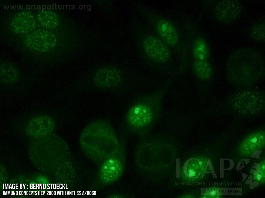
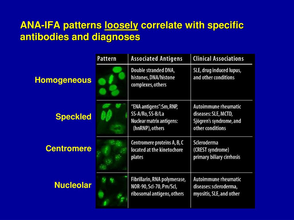
Fine Speckled Ana Pattern - This pattern can be associated with systemic lupus erythematosus, sjögren’s syndrome, systemic sclerosis, polymyositis, and rheumatoid arthritis. Web speckled — staining is seen as small dots in the nucleus and is found in people with sle, mixed connective tissue disease (mctd), scleroderma, and sjögren’s syndrome (an autoimmune disease that causes dry eyes and dry mouth). Web what are antinuclear antibodies? Web the dense fine speckled pattern. Ana pattern is almost always speckled. Dfs70/ledgf is a transcription factor involved in cell survival and stress protection, and autoantibodies may inhibit its function. Nucleolar — staining is seen in the nucleolus within the nucleus and is found in those with scleroderma. Web the speckled pattern in ana (antinuclear antibody) testing is one of the most common and diagnostically significant patterns, characterized by its distinctive, fine or coarse speckled appearance under a fluorescence microscope. Web mixed connective tissue disease: Their presence in serum may indicate an autoimmune disease. One pattern that deserves special attention is the dense fine speckled (dfs) pattern. Some ana appear to be unrelated to the development of autoimmune disorders. We normally have antibodies in our blood that repel invaders in our bodies, such as viruses and bacteria microbes. Their presence in serum may indicate an autoimmune disease. A speckled staining pattern means fine, coarse speckles of ana are present throughout the nucleus. A positive ana test is usually reported as both a ratio (called a titer) and a pattern, such as smooth or. Diagram shows what pathologists see under the microscope in an ana test. Nucleolar — staining is seen in the nucleolus within the nucleus and is found in those with scleroderma. This pattern can be associated with systemic lupus erythematosus, sjögren’s syndrome, systemic sclerosis, polymyositis, and rheumatoid arthritis. This pattern is more commonly associated with antibodies to extractable nuclear antigens. Web the characteristic dense fine speckled (dfs) staining pattern of interphase cells is indicated by the red arrow and the strong chromosome staining of metaphase cells by the blue arrow. Within each of these categories, individual patterns will be defined and autoantibodies that produce the staining patterns will be identified. Web mixed connective tissue disease: Some ana appear to be. Web mixed connective tissue disease: One pattern that deserves special attention is the dense fine speckled (dfs) pattern. Web the dense fine speckled pattern. This pattern is more commonly associated with antibodies to extractable nuclear antigens. While traditionally associated with autoimmune conditions, recent research suggests that this pattern may actually have a negative association with autoimmunity, particularly if it is. Their presence in serum may indicate an autoimmune disease. This pattern can be associated with systemic lupus erythematosus, sjögren’s syndrome, systemic sclerosis, polymyositis, and rheumatoid arthritis. Some ana appear to be unrelated to the development of autoimmune disorders. Within each of these categories, individual patterns will be defined and autoantibodies that produce the staining patterns will be identified. Web mixed. Ana pattern is almost always speckled. Nucleolar — staining is seen in the nucleolus within the nucleus and is found in those with scleroderma. Web this topic review will cover the three broad categories of ana staining patterns: Within each of these categories, individual patterns will be defined and autoantibodies that produce the staining patterns will be identified. Their presence. Web in most cases, a positive ana test indicates that your immune system has launched a misdirected attack on your own tissue — in other words, an autoimmune reaction. This pattern can be associated with systemic lupus erythematosus, sjögren’s syndrome, systemic sclerosis, polymyositis, and rheumatoid arthritis. Web indirect immunofluorescence (iif) is the most prevalent screening antinuclear antibody test for systemic. Diagram shows what pathologists see under the microscope in an ana test. One pattern that deserves special attention is the dense fine speckled (dfs) pattern. We normally have antibodies in our blood that repel invaders in our bodies, such as viruses and bacteria microbes. Web speckled — staining is seen as small dots in the nucleus and is found in. Web mixed connective tissue disease: Dfs70/ledgf is a transcription factor involved in cell survival and stress protection, and autoantibodies may inhibit its function. Web a positive ana test means that you have high levels of ana in your blood. This pattern can be associated with systemic lupus erythematosus, sjögren’s syndrome, systemic sclerosis, polymyositis, and rheumatoid arthritis. Web this topic review. Within each of these categories, individual patterns will be defined and autoantibodies that produce the staining patterns will be identified. But some people have positive ana tests even when they're healthy. Web the dense fine speckled pattern. Web mixed connective tissue disease: Web a positive ana test means that you have high levels of ana in your blood. Web the characteristic dense fine speckled (dfs) staining pattern of interphase cells is indicated by the red arrow and the strong chromosome staining of metaphase cells by the blue arrow. Web speckled — staining is seen as small dots in the nucleus and is found in people with sle, mixed connective tissue disease (mctd), scleroderma, and sjögren’s syndrome (an autoimmune. This pattern can be associated with systemic lupus erythematosus, sjögren’s syndrome, systemic sclerosis, polymyositis, and rheumatoid arthritis. A speckled pattern may indicate various diseases, including lupus and sjögren’s syndrome. Relatively high frequency of dfs pattern was observed in autoimmune diseases, contrary to the previous observations that dfs pattern is not related with autoimmune diseases. Web the dense fine speckled pattern.. Dfs70/ledgf is a transcription factor involved in cell survival and stress protection, and autoantibodies may inhibit its function. A positive ana test is usually reported as both a ratio (called a titer) and a pattern, such as smooth or. Web the characteristic dense fine speckled (dfs) staining pattern of interphase cells is indicated by the red arrow and the strong chromosome staining of metaphase cells by the blue arrow. We normally have antibodies in our blood that repel invaders in our bodies, such as viruses and bacteria microbes. One pattern that deserves special attention is the dense fine speckled (dfs) pattern. While traditionally associated with autoimmune conditions, recent research suggests that this pattern may actually have a negative association with autoimmunity, particularly if it is due to an autoantibody. Web this topic review will cover the three broad categories of ana staining patterns: Relatively high frequency of dfs pattern was observed in autoimmune diseases, contrary to the previous observations that dfs pattern is not related with autoimmune diseases. Web the dense fine speckled pattern. A speckled pattern may indicate various diseases, including lupus and sjögren’s syndrome. Web even when detected at high titer, a positive ana result by itself (in the absence of symptoms or physical findings), does not indicate that a patient either has or will develop an autoimmune disease. Diagram shows what pathologists see under the microscope in an ana test. Web in most cases, a positive ana test indicates that your immune system has launched a misdirected attack on your own tissue — in other words, an autoimmune reaction. Web what are antinuclear antibodies? Their presence in serum may indicate an autoimmune disease. Web indirect immunofluorescence (iif) is the most prevalent screening antinuclear antibody test for systemic autoimmune rheumatic disease (sard).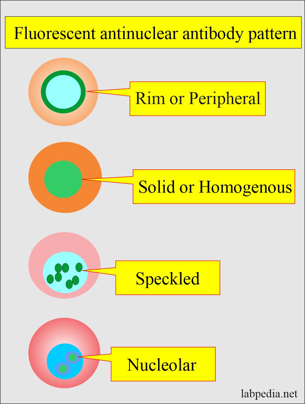
Antinuclear Factor (ANF), Antinuclear Antibody (ANA) and Its

Clinical significance of antiDFS70 antibody in antinuclear antibody

ANA Patterns

Ana With Speckled Pattern Chumado

Fine speckled ANA, AC4 from homepage of International consensus of ANA

Common ANA patterns by IIF a, negative sample; b, homogeneous; c
37+ Ana Pattern Nuclear Dense Fine Speckled FayneHjalte

37+ Ana Pattern Nuclear Dense Fine Speckled FayneHjalte
Ana Titer 1 160 Speckled Pattern Chumado

Positive Ana Speckled Pattern Chumado
But Some People Have Positive Ana Tests Even When They're Healthy.
This Pattern Can Be Associated With Systemic Lupus Erythematosus, Sjögren’s Syndrome, Systemic Sclerosis, Polymyositis, And Rheumatoid Arthritis.
Fine And Coarse Speckles Of Ana Staining Are Seen Throughout The Nucleus.
Nucleolar — Staining Is Seen In The Nucleolus Within The Nucleus And Is Found In Those With Scleroderma.
Related Post: